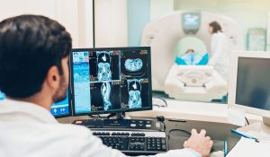
 Body imaging is the subspecialty of radiology utilizing advanced techniques to evaluate the organs in the chest, abdomen and pelvis. After four years of general radiology education, each of our dedicated body imaging specialists completed an additional year of training (fellowship), to develop expertise in advanced cross-sectional imaging. They have brought their interpretation skills and the newest imaging techniques to South Florida, from their experiences at prestigious institutions such as Johns Hopkins, Yale, Massachusetts General Hospital and the NYU Medical Center.
We do not believe in a “one scan fits all” approach to radiology. Each patient has his or her own symptoms and medical history. At RAH, we choose from a variety of sophisticated imaging protocols, adjusting our scanning parameters to tailor your MRI or CT scan to your specific clinical problem. Our state-of-the-art scanners have been programmed by our own radiologists to provide these customized studies.
We work closely with our clinical colleagues, including surgeons, oncologists, emergency department physicians, gastroenterologists, pulmonologists, urologists, and general internists. We are proud of the reputation of excellence that we have developed by consistently providing outstanding images and accurate specialized interpretations to our referring physicians.
Procedures
In the field of Body Imaging some of our more popular procedures are:
- CT scans of the abdomen and pelvis – can evaluate generalized abdominal pain, but also perform specialized scans tailored to image individual organs such as the liver, pancreas, kidneys, adrenal glands, and intestines.
- CT scan of the chest – including lung cancer screening, lung nodule follow-up and high-resolution scanning. Often combined with a scan of the abdomen and pelvis to evaluate and stage many types of cancer.
- MRI of the abdomen – including specialized imaging protocols for the liver, pancreas and biliary system (MRCP), kidneys and adrenal glands.
- MRI of the pelvis – to image the ovaries and uterus, evaluating pelvic pain, pelvic masses, ovarian cysts and uterine fibroids.
- Cardiac imaging – including coronary calcium scoring and coronary artery CT angiography (CTA), using high resolution CT Scanners across the Memorial Healthcare System (Source: RadiologyInfo.org).
Physicians
- Karen Ayres, MD
- Jordan Ditchek, MD
- Margaret Gallegos, MD
- David Hodge, MD
- Charles Jacobs, MD
- Matthew Lee Hoffman, MD
- Jeffrey A. Katz, MD
- Andrew Hai Mai, MD
- Roberto Maldonado, MD
- Barbara Raphael, MD
- Raymond Ramzi, MD
- Mark Schwimmer, MD
- Scott Skorupa, MD